The scales below were pulled from an orange and black Clarion Angelfish that probably looked like the picture below when alive:

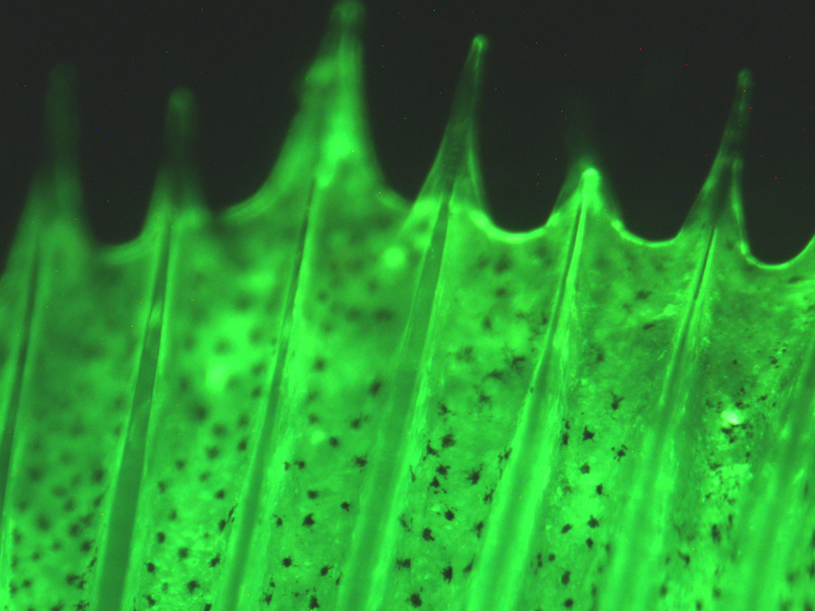
These are all pictures of the black scales. Most of the scale looks translucent. This is because most of the coloration is at the very end of the scale (far left in picture). When the magnification is increased, there are black dots that become more distinguishable. Is it these small dots alone that give the fish its black color? Is the coloration due to pigment or the structure of the scale? These are questions we seek to answer. But first, at least a couple of black scales had to be photographed.
Note: If you click on the picture, it will open up in its actual size, allowing you to see the high resolution and detail that the microscope camera is capable of.
Black Scale One Face Up (Some Overlap)
Full Scale View:


Detail of Tips, note the black dots especially in the second picture


Bottom edge that is usually under other scales on the fish


A small overlapping scale with lots of visible black dots
(from what Caitlin told me, an overlapping scale isn't as common as you'd think)


Middle of the Scale


Higher Magnification Center of Scale


Black Scale One Underside


Well, if you made it to the end (or just skipped on down here), then I'd first like to congradulate you. When a phase of this project is nearing conclusion, tens and tens more photos will have to be looked through, compared, analyzed, etc. to give an accurate photographic analysis of whether or not fish scale iridescence and coloration is due to pigmentation or structure. It is interesting to note that this is only one set of photos for only one scale. There are always more than 2 sets of photos per scale color according to location on the fish, which means there are anywhere from 6 to 10 photo-sets per fish, and this was Fish #7. Not to mention that I'm not counting the photo sets of digested scales, which these photos will be juxtaposed with.
In addition, this is one of several photo sets I took of the Clarion Angelfish scales.